5 Common Orthopedic Problems:
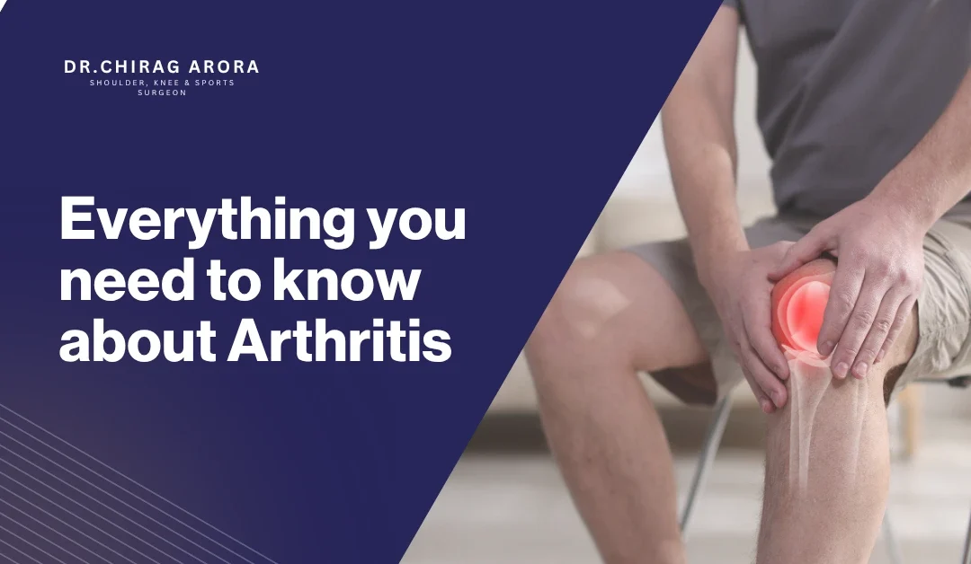
- Osteoarthritis (Wear and Tear of Joints)
Osteoarthritis is a common problem with joints that older adults get. It happens when the cartilage that protects our bones wears down over time.
Symptoms of osteoarthritis include:
- Joint pain in the knees, hips, and hands
- Feeling stiff in the morning
- Swelling in the joints
- Trouble moving
Causes of osteoarthritis are:
- Getting older
- Using our joints too much
- Being overweight
- Hurting our joints in the past
Treatment for Osteoarthritis:
- Watch our weight
- Do physiotherapy
- Take medicine to relieve pain
In some cases, we can even get a new joint. If we take care of osteoarthritis early, it can really help slow it down.
- Osteoporosis (Weak Bones)
Osteoporosis makes our bones weak. This means we can break them easily even if we just fall down or get hurt.
Symptoms of Osteoporosis include:
- Breaking bones a lot
- Having back pain
- Getting shorter over time
- Having posture
Causes of Osteoporosis are:
- Not having enough calcium and vitamin D
- Hormonal changes, especially in women after menopause
- Not being active
- Getting older
Treatment of Osteoporosis we can:
- Take calcium and vitamin D supplements
- Do exercises that make our bones stronger
- Take medicine to make our bones stronger
- Change our lifestyle
Osteoporosis is sometimes called a “silent disease” because we do not know we have it until we break a bone.
- Cervical Spondylosis (Neck Pain)
Cervical spondylosis is when the bones and discs in our neck wear down as we age.
Symptoms of spondylosis include:
- Having a stiff neck
- Getting headaches, especially at the back of our head
- Having pain in our shoulders
- Feeling tingling in our arms or hands
Causes of spondylosis are:
- Using screens for too long
- Having bad posture
- Our spinal discs are getting older
Treatment of Cervical Spondylosis we can:
- Do neck exercises
- Do physiotherapy
- Take medicine to relieve pain
Change how we live and work to reduce strain on our neck. If we sit up straight while we work or use our phones it can really help reduce the symptoms.
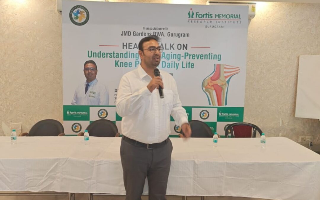
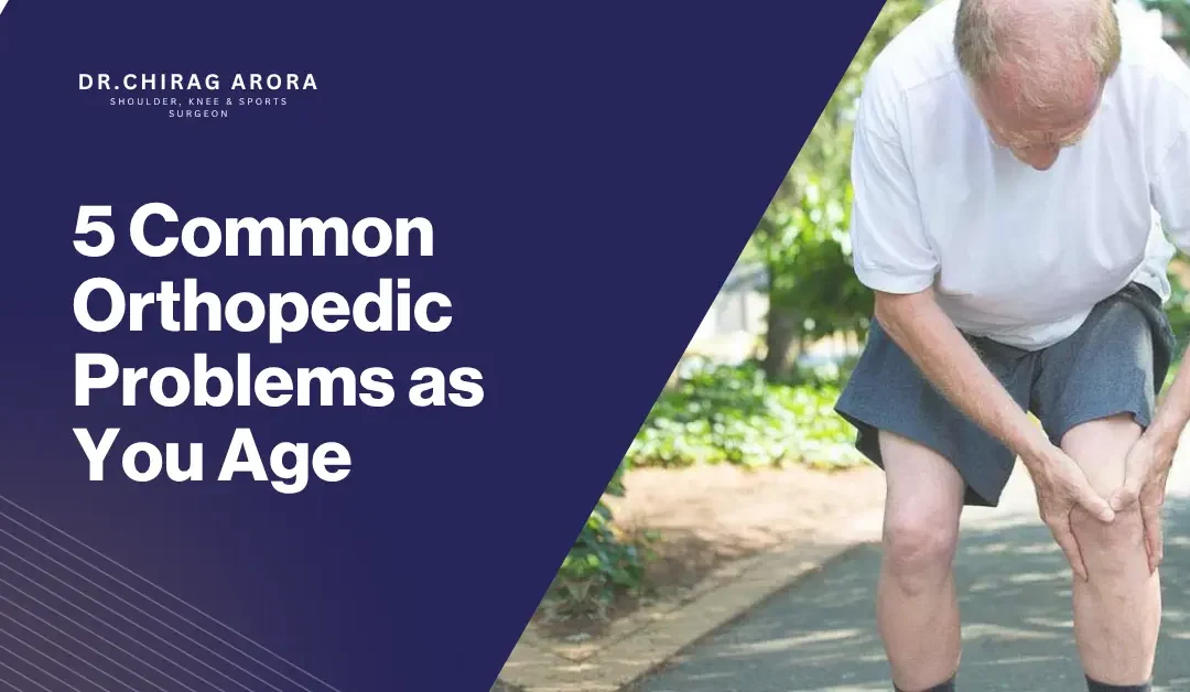
- Knee Pain & Degenerative Joint Disease
Knee problems are very common as we age, especially because the cartilage in our knees wears down.
Symptoms of Knee Problems include:
- Having pain when we walk or climb stairs
- Having swollen knees
- Hearing a cracking sound in our joints
- Having trouble standing for a time
Causes of Knee Problems are:
- Getting older and wearing down our joints
- Being overweight
- Hurting our knees in the past
- Not having strong muscles
To treat Knee Problems we can:
- Do physiotherapy and exercises to strengthen our muscles
- Use knee braces
- Lose weight
In cases, we may also have to get knee replacement surgery. If we have strong thigh muscles it can really help support our knees and reduce pain.
- Lower Back Pain (Lumbar Spondylosis / Disc Problems)
Lower back pain is one of the common orthopedic problems that adults have.
Symptoms of Back Pain include:
- Having constant or recurring back pain
- Having pain when we bend or lift things
- Having a stiff lower back
- Having pain that goes down our legs
Causes of lower back pain are:
- Having spinal discs that are worn out or slipped
- Having bad posture
- Having weak core muscles
- Sitting for too long
To treat lower back pain we can:
- Do physiotherapy
- Do exercises to strengthen our core muscles
- Manage our pain
- Change our lifestyle
If we ignore back pain, it can lead to big problems with moving around.